New Jersey, USA


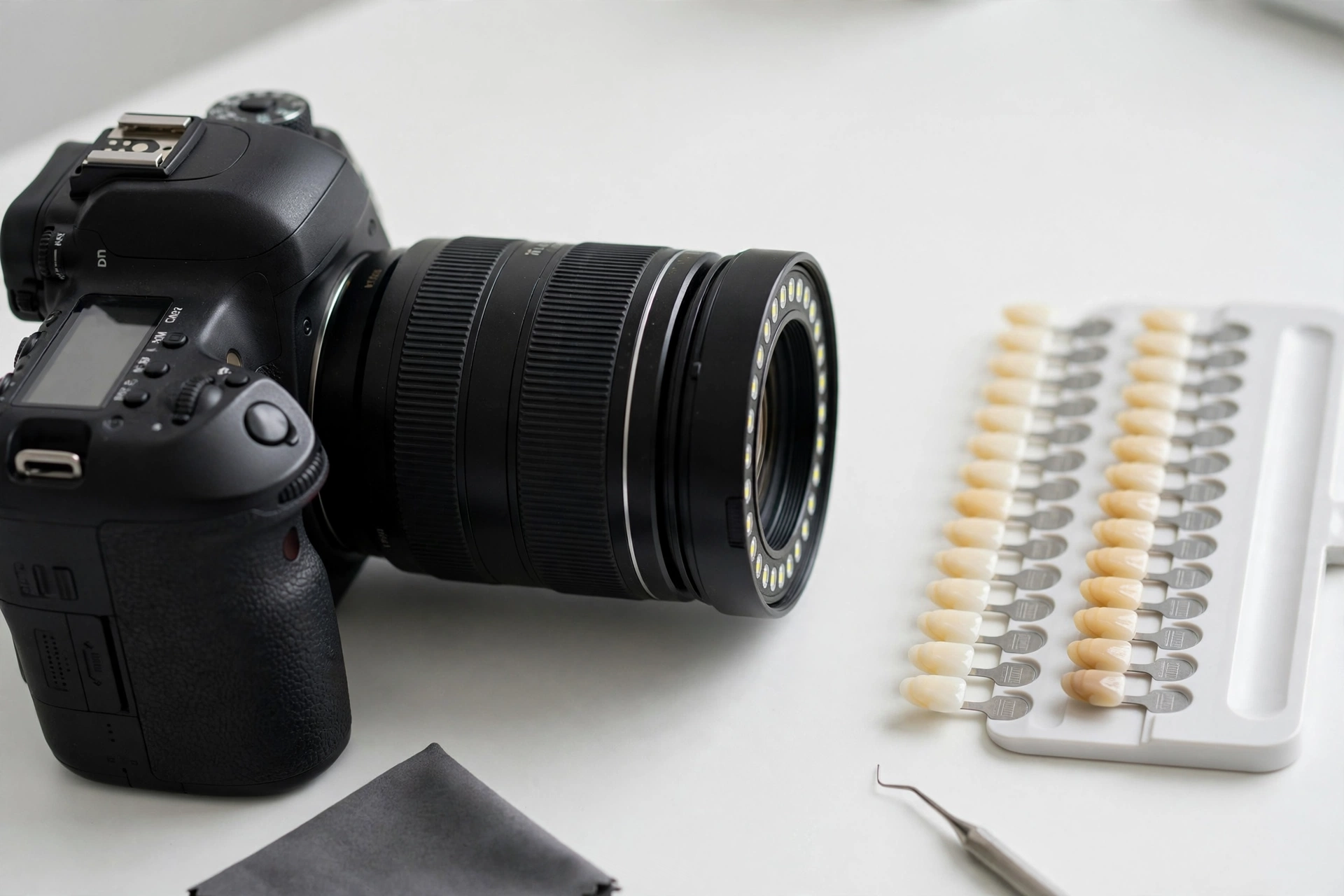
Successful dental restorations depend on more than technical expertise and advanced laboratory equipment. Clear communication between dentists and dental technicians is equally important. One of the most effective ways to transfer visual clinical information is through high-quality dental photography.
Photographs can provide the laboratory with details that may not be fully communicated through written instructions, digital impressions, or shade information alone. When captured correctly, they help technicians understand the patient’s existing dentition, aesthetic expectations, tooth characteristics, and clinical situation.
Why Is Dental Photography Important for Dental Laboratories?
Dental technicians often design restorations based on information received from the dental practice. Although digital scans provide highly detailed three-dimensional data, they may not fully communicate factors such as facial proportions, soft-tissue appearance, surface texture, or the visual relationship between teeth and the surrounding structures.
Dental photography can complement digital records by providing additional visual information about the patient.
This is particularly valuable for:
- Anterior aesthetic restorations
- Veneers and crowns
- Full-mouth rehabilitation
- Implant-supported restorations
- Smile design
- Complex shade-matching cases
- Highly aesthetic ceramic restorations
What Information Can Dental Photographs Provide?
A well-planned photographic series can communicate several important clinical details to the laboratory.
Tooth Shape and Proportions
Photographs can help technicians evaluate the overall shape, width-to-length ratio, incisal edge position, and relationship between adjacent teeth.
These details are particularly important when fabricating anterior restorations where small differences in morphology can affect the final appearance.
Shade and Characterization
Shade communication can be challenging because tooth color is influenced by lighting, camera settings, surrounding colors, and the photography environment.
Standardized photographs can provide additional visual references for the technician when assessing hue, chroma, value, translucency, and surface characterization.
However, photographs should complement rather than replace proper clinical shade-taking procedures.
Surface Texture
Natural teeth are rarely completely smooth. Developmental features, enamel texture, subtle grooves, and surface irregularities contribute to their appearance.
High-quality close-up photographs can help technicians reproduce these characteristics more naturally when designing ceramic restorations.
Gingival and Soft-Tissue Information
The relationship between the restoration and surrounding soft tissue is especially important in aesthetic and implant cases.
Photographs can show gingival contours, papillae, tissue levels, and asymmetries that may influence restoration design.
How Photography Improves Dentist–Technician Communication
Written laboratory prescriptions can describe the requested restoration, but photographs can provide context that words alone cannot easily convey.
For example, a dentist may request a natural-looking anterior crown. A photographic record can help the technician understand what “natural” means in that specific case by showing the patient’s existing teeth, facial characteristics, smile line, and surrounding structures.
This additional information can reduce ambiguity and support more predictable collaboration.
Dental Photography in Digital Workflows
Modern dental laboratories increasingly work within digital workflows that combine intraoral scanning, CAD design, digital photography, and other clinical records.
Photographs can be integrated into digital case documentation and used alongside scans during the design process.
When the laboratory receives both three-dimensional digital data and standardized photographic references, technicians can evaluate the case from multiple perspectives.
This combination is especially useful for complex aesthetic cases where shape, color, texture, and facial harmony must be considered together.
What Makes a Good Dental Photograph for Laboratory Use?
Not every clinical photograph provides the same level of information. For laboratory communication, consistency is important.
Useful photographs should generally have:
- Consistent lighting
- Accurate focus
- Appropriate exposure
- Minimal reflections
- Correct positioning
- Clean retraction where necessary
- Adequate magnification
- A neutral and controlled background when appropriate
A standardized photographic protocol makes it easier for technicians to compare images and interpret clinical details accurately.
Common Photography Mistakes
Poor photographic documentation can create confusion rather than improve communication.
Common problems include:
- Incorrect white balance
- Excessive shadows
- Overexposure or underexposure
- Blurry images
- Inconsistent camera angles
- Inadequate retraction
- Missing occlusal or lateral views
- Using only one photograph for a complex case
Providing a complete and consistent photographic series is generally more useful than sending a single close-up image.
Which Photographs Should Be Sent to the Laboratory?
The exact photographic protocol depends on the case. However, a comprehensive record may include:
Full-face photographs:
Useful for evaluating facial proportions, smile dynamics, and overall aesthetic relationships.
Smile photographs:
Help the technician understand tooth display, incisal position, smile line, and symmetry.
Retracted frontal photographs:
Provide a detailed view of the anterior teeth and gingival architecture.
Lateral photographs:
Help demonstrate tooth position, contours, and relationships between the anterior and posterior segments.
Occlusal photographs:
Provide additional information about arch form, tooth positioning, and existing anatomy.
Close-up photographs:
Useful for examining shade, translucency, characterization, and surface texture.
The Laboratory’s Role in Photographic Communication
Dental technicians should also understand how to interpret photographic information rather than simply receive images as attachments.
When photographs are reviewed together with scans, prescriptions, shade information, and clinical notes, the laboratory can build a more complete understanding of the case.
If important information is missing or unclear, communication between the technician and dentist should take place before fabrication begins.
Dental Photography and Remake Prevention
Restoration remakes can occur when the laboratory does not have sufficient information to reproduce the intended clinical result.
While photography cannot eliminate every potential source of error, comprehensive visual documentation can reduce uncertainty in cases where aesthetics and morphology are particularly important.
Better communication before manufacturing may also reduce unnecessary modifications and chairside adjustments.
Conclusion
Accurate laboratory communication requires more than sending a digital impression and a written prescription. Dental photography adds valuable visual information that can help technicians understand tooth morphology, shade characteristics, soft-tissue relationships, facial aesthetics, and the patient’s existing condition.
When standardized photographs are combined with digital scans and clear clinical instructions, they create a stronger communication pathway between the dental practice and laboratory.
For modern restorative dentistry, dental photography is therefore an important part of comprehensive case documentation and predictable laboratory collaboration.
Frequently Asked Questions
Why should dentists send photographs to the dental laboratory?
Photographs provide visual information about tooth shape, shade, texture, soft tissues, smile characteristics, and facial relationships that may not be fully visible in digital scans.
Are photographs necessary for every dental laboratory case?
Not necessarily. The need depends on the complexity and aesthetic requirements of the restoration. They are particularly valuable for anterior, implant, and full-mouth aesthetic cases.
Can photographs replace a digital impression?
No. Photographs and digital impressions provide different types of information and are most effective when used together.
How can dentists improve photographic communication with technicians?
Using a consistent photographic protocol, appropriate lighting, accurate exposure, multiple clinical views, and clear case instructions can significantly improve the quality of communication.
Do dental photographs help with shade matching?
They can provide useful visual references for shade and characterization, but standardized clinical shade-taking remains important because photographic color can be affected by lighting and camera settings.